
The Female breasts, also known as the ‘mammary glands,’ are the accessory reproductive organs. Although they are not directly involved in pregnancy or childbirth, they do play an important role in the baby’s growth after birth.
Situated on the chest, breasts are present in both males and females but in males, the breast structures do not grow and remain rudimentary.
During childhood, a young girl’s breasts are similar to those of a young boy, consisting of a simple system of ducts.
Breasts begin to develop at around the age of 9 – 10 years, just before the first menstrual period, under the influence of growth hormones, adrenal cortical hormones as well as estrogen secreted by the ovaries.
Structure of the Breast
Breasts are pear-shaped structures composed of glandular tissue, fibrous tissue, fatty tissue and blood vessels.
The size of the breasts varies among women. The tissue composition of the breasts determines the firmness or density of the breasts. This too can vary among different women.
The shape, size, and weight of the breasts changes in women in different stages of life like puberty, pregnancy, breastfeeding, and menopause.
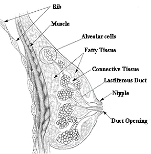
An adult female breast consists of around 15- 20 lobes of glandular tissue. Each lobe is made up of several lobules that radiate around the nipple. Fat deposited around the lobules give the breasts their typical rounded shape.
Each of these lobules is composed of a group of specialized cells known as the alveoli. The alveoli are capable of producing milk from blood. Milk is drained from the alveoli by small ducts, which collectively form large excretory ducts called lactiferous ducts. . A single lactiferous duct drains each lobule.
These lactiferous ducts, supported by the dense connective tissues, converge towards the center of the breasts to form reservoirs of milk known as the ampulla (also called lactiferous sinus). A single duct arises from each ampulla to open into the tip of the nipple. When the baby suckles, the milk from the ampulla is released through these lactiferous ducts.
Various arteries, veins, and lymph nodes provide blood and lymph supply to the breasts. The network of connective tissue and ligaments provide support and shape to the glands.
The breast is anchored to the pectoral muscles on the chest wall by strands of fibrous tissue. Loosening of these fibrous strands can cause the breasts to sag In older women. Thin strands of fibrous tissue also attach the breast to the overlying skin.
Functions of the Breast
- The primary function of the breasts is to provide nourishment to the infant through breastfeeding. When an infant sucks on the mother’s nipples, the mother’s brain secretes oxytocin. Increase in the level of oxytocin triggers the contraction of cells surrounding the alveoli and causes the release of milk through the duct to the nipples.
- In addition to their primary function of providing nutrients to the infant, breasts also have a sexual role. Breasts, and especially the nipples, are an erogenous zone and respond to sexual stimuli.
- Presence of breasts also contribute to society’s image of a typical female body and is a measure of sexual attractiveness in women.
Size of the Breast
The amount of fat surrounding the lobules greatly influences the size of the breast. As a result, a larger woman may have larger breasts than a thinner woman.
Because the ability of the breast to secrete milk is determined by its lobules rather than its fat, the size of the breast has no relation to the amount of milk the breast can produce while breastfeeding.
The Areola
The areola is a 2 – 3 cm pigmented area located in the centre of the breast. It has a number of sebaceous and sweat glands, which keep it moist and supple.
The areola is provided with modified sebaceous glands, called Montgomery’s glands or tubercles that run along the edge of the areola and on the nipple itself. These glands secrete an oily fluid that helps lubricate and protect the nipple during lactation. Montgomery’s tubercles range from 4 to 28 on each areola. They become larger and more prominent during pregnancy.
The fluid released by the Montgomery’s tubercles contains different volatile compounds which stimulate the baby’s appetite during breastfeeding.
The Nipple
The nipple is a conical protrusion present at the center of the breast. It contains the 15 to 20 lactiferous ducts and their openings. It is rich in nerve endings, making it a highly sensitive organ for many women. It contains erectile muscles that causes the nipple to become erect when stimulated. Although the breast contains a large amount of fat, there is no fat in the nipples or underneath the areola.
Breast Changes during Puberty:
During puberty, the female body experiences a surge in hormones, particularly estrogen, that stimulates the growth of the breasts. As a result, the breasts begin to develop from small buds into full, mature breasts. The nipples and the areola become more prominent and the size and color of the areola changes as the breasts develop. The areola may become larger and darker, and the skin may become more sensitive.
This process can take several years to complete and can result in significant changes in the size and shape of the breasts.
Breast Changes During the Menstrual Cycle
The level of oestrogen and progesterone secreted during the menstrual cycle causes cyclic changes in breast tissue.
The level of oestrogen rises early in the menstrual cycle, causing some enlargement of the breast ducts. The Progesterone level rises in the latter half of the menstrual cycle and causes alveolar cell enlargement.
These changes may cause breast enlargement and tenderness just before the menstrual period. Progesterone may cause fluid retention in the premenstrual week. This is more common in women suffering from premenstrual syndrome (PMS) and a condition known as fibrocystic breast disease.
When the menstrual period begins and hormonal levels fall, the tenderness and enlargement decreases.
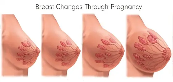
Breast Changes During Pregnancy
During pregnancy, the breasts undergo many changes as they prepare for breastfeeding. Some of these changes include:
Growth: During the early stages of pregnancy, the pituitary gland secretes a hormone called prolactin. Prolactin causes growth and enlargement of both the lobules as well as the lactiferous ducts. The breasts typically begin to grow larger during the first trimester of pregnancy. They continue to grow throughout pregnancy due to increased blood flow to the breast tissue. Fine veins on the breast’s surface may become visible.
Tenderness: The breasts may become sore, tender, or sensitive to the touch due to hormonal changes and growth of the breast tissue. The nipples and areolas can become more sensitive and painful to touch. This tenderness is often most noticeable in the first trimester and can last throughout pregnancy.
Darkening of the Areola: The areola, the dark area surrounding the nipple, may become darker and more pronounced during pregnancy due to increased production of melanin, the pigment that gives color to the skin, and increased blood flow. A pigmented area may develop around the areola – this is known as the secondary areola.
Nipple Changes: The nipples may become more sensitive and may become more prominent due to hormonal changes and increased blood flow.
Lumps and Bumps: Some women may develop lumps or bumps in the breast tissue during pregnancy due to growth of the alveolar cells in the lobules. These lumps are typically harmless and are caused by changes in the ducts and glands in the breast.
Colostrum: During the third trimester of pregnancy, the breasts may secrete colostrum, a thin whitish liquid. Colostrum secretion is usually induced by squeezing the breasts, but it can also happen on its own.
After Childbirth: Breasts may grow in size even more after childbirth when breastfeeding begins. Breast engorgement and abscesses are possible complications during this time.
Overall, these changes in the breast during pregnancy are normal and a natural part of preparing for lactation.
Breast Changes after Menopause:
After menopause, the hormonal changes that occur can result in a reduction in breast size and firmness. The mammary glands begin to shrink, and the amount of fatty tissue in the breasts decreases, leading to a loss of fullness. The skin can also become less elastic, leading to sagging or drooping of the breasts. The skin of the areola and nipples may become lighter, and the size may shrink.
Breast Cancer
Breast cancer can develop at any time during a woman’s reproductive life, though some types are more common after menopause. Some common symptoms of breast cancer include a lump in the breast, changes in the size or shape of the breast, and changes in the skin or nipples . Mammograms should be performed every two years, especially in women at high risk of breast cancer.

Read Questions and Answers on Pregnancy and Gynecology
Natural PCOS Solutions: Control Symptoms, Manage Irregular Periods, and Reduce Excessive Hair Growth
Tackling PCOS Symptoms: A Patient’s Journey with Dr. Madhumita Mazumdar Polycystic Ovary Syndrome (PCOS) is a common hormonal disorder that can significantly impact young women’s

Early Pregnancy Bleeding: Causes, Symptoms and Treatment
Early Pregnancy Bleeding can be due to benign causes like implantation bleeding or due to serious causes like miscarriage or ectopic pregnancy.
HPV: 10 Important Facts
Human papillomavirus (HPV) is a sexually transmitted infection (STI) that can cause warts and cancers. There are more than 100 strains of HPV, some more dangerous than the others.
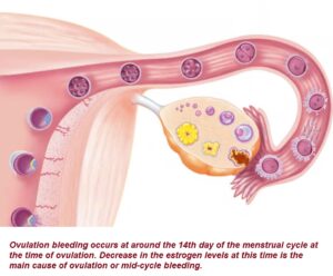
Ovulation Bleeding: Symptoms, Causes and Treatment
Ovulation bleeding, also known as mid-cycle bleeding occurs at around the 14th day of the menstrual cycle.

Abdomen Pain in Early Pregnancy: Causes and Treatment
Pain in the abdomen is common in early pregnancy. These can be due to benign causes like UTI or intestinal gas. But miscarriage needs to be ruled out.

Uterine Fibroids: A Patient’s Journey to Diagnosis and Treatment
Uterine fibroids, also called ‘Myomas’, are non-cancerous growths that develop in the muscles of the uterus, affecting many women during their reproductive…