
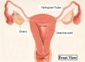
Uterus
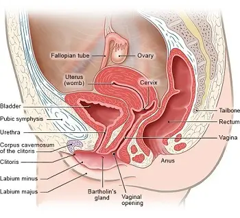
The uterus is a pear-shaped, muscular organ located deep within the pelvic cavity, between the bladder in front, and the rectum behind it, well protected by the pelvic bones.
It is the main organ of the female internal reproductive system. Its primary function is to provide a supportive and nourishing environment for a developing fetus during pregnancy.
From the front, it resembles an inverted triangle, with the broad base serving as the roof and the narrow apex at the lower end. This lower end, which is also known as the cervix, opens into the vagina. The two angles on the two ends of the triangle’s base are connected to the two Fallopian tubes.
The uterus is held in place by various ligaments, including the broad ligament and the round ligament.
At its thickest point, the adult uterus is about 8cm long and 5cm wide. Its length ranges from 2cm to 5cm before menstruation begins. The weight ranges from 50 to 80 grams. The uterus can only hold about 5ml of fluid when not pregnant.
The uterus is typically about the size of a fist in a non-pregnant woman, but it can expand greatly during pregnancy. When a woman becomes pregnant, her uterine muscles expand in both number and size (up to 500 times their normal size). The uterus is the organ that receives, implants, retains, and feeds the foetus until it is fully developed. It grows gradually from the first trimester to the second trimester and finally to the end of the third trimester. During labour and childbirth, it contracts and expels the foetus.
After the menopause, the uterus becomes considerably smaller in size due to lower levels of estrogen and progesterone.
Ovaries
The ovaries are two small, almond-shaped organs located on either side of the uterus. They are the female sex gonads and produce the eggs, or ova, that are released during ovulation. They also produce hormones, such as estrogen and progesterone, that regulate the menstrual cycle and support pregnancy.
The ovaries are pinkish-white in color and roughly oval in shape, measuring approximately 3cm in length, 2cm in breadth, and 1cm in thickness. Each ovary has a thick outer lining called the ‘cortex’ and a thin inner layer called the ‘medulla.’
The ovaries are held in place by various ligaments, including the ovarian ligament, which attaches the ovary to the uterus, and the suspensory ligament, which helps to support the ovary within the pelvic cavity.
The cortex contains numerous ‘Graafian follicles’ at various stages of development during the reproductive life, i.e. from puberty to menopause. Every month, a Graafian follicle matures and releases an ovum from one of the ovaries. This is referred to as ‘ovulation.’ Only about 400 follicles mature in a woman’s lifetime.
During puberty, the ovaries will grow in response to hormonal signals and begin to release eggs. Over the course of the menstrual cycle, the ovaries will also change in size and shape, as follicles mature and eggs are released during ovulation.
Ovaries play a crucial role in reproductive life. The ovum (egg) develops in the ovaries. Ovulation is the process by which a mature egg is released from the ovary and travels down the fallopian tube, where it may be fertilized by sperm. The ovaries produce hormones, including estrogen and progesterone, which help to regulate the menstrual cycle and maintain fertility.
Menopause occurs when the number of follicles in a woman’s ovaries falls below a critical level. The ovaries shrink and turn whitish in color. Their hormone-producing capabilities decline leading to lower lower levels of estrogen and progesterone in the blood.

The Fallopian tubes
The fallopian tubes, also known as the ‘Oviducts,’ are a key part of the female reproductive system and play an important role in fertility and pregnancy.
They are located on either side of the uterus, one on each side. They extend like arms from the uterus, reaching for the ovaries, which are located one on each side of the uterus. Each tube has two openings. One of the openings leads to the uterus. The other opening is larger and wider, with a number of finger-like projections called ‘fimbriae’ all around it.
The fimbriae are located near the ovary on the same side and pick up the ovum when it is released from the ovary (‘ovulation’). The inner lining of the tube contains cells with microscopic hairs called cilia, which help in propelling the ovum towards the uterus.
Each tube is about 10cm long. The width varies along the length, with the ovarian side being wider and the uterine side being thinner but more muscular. The ampulla, its widest part, is located next to the fimbria and is important because sperm fertilization of the ovum usually occurs in this region.
The fallopian tubes are held in place by various ligaments and muscles within the pelvic cavity. They are positioned in such a way that they can easily pick up an egg that has been released from the ovary and transport it to the uterus.
The tubes are the pathways through which the egg travels from the ovaries to the uterus. If fertilization occurs, it typically takes place within the fallopian tubes.
The fallopian tubes are susceptible to various infections such as pelvic inflammatory disease (PID) and blockages, which can affect fertility and increase the risk of complications during pregnancy.

Vagina
The vagina is a hollow, elastic, fibromuscular structure that extends upwards and backwards from the vulva.
It connects the uterus to the body’s external surface via the vaginal opening in the vulva. The uterine cervix protrudes into the vagina at its upper end.
It is approximately 2.5 cm wide and 7 to 9 cm long has a slightly curved shape. The walls of the vagina are made up of several layers of muscles and are moist and flexible, allowing them to expand and contract in response to sexual arousal or childbirth.
The vagina is positioned within the pelvic cavity, between the bladder in front and the rectum behind it. The external opening of the vagina, known as the introitus, is located in the vestibule of the vulva, between the urethral opening and the anus.
The vagina is related to many important structures. The Bartholin’s glands are positioned on either side of the vaginal opening – they secrete a natural lubricant to help keep the vagina moist. The G-spot, is an area located on the front wall of the vagina and is associated with sexual pleasure.
The vaginal organ is the female organ for sexual intercourse and forms the birth canal during childbirth. During the menstrual period, it is also the opening through which the menstrual blood flows out.
The vagina plays several important roles. It is the passageway for menstrual blood to flow from the uterus to the vulva, it is the birth canal for a baby during childbirth, and it is the female organ for sexual intercourse.
The vagina is at risk of developing various infections like yeast infections, bacterial vaginosis, and sexually transmitted infections (STIs). A normal vaginal ph and a proper hygiene is crucial for maintaining vaginal health.
The Ligaments
The ligaments are strong, fibrous cords that connect and support the uterus, ovaries, and other reproductive organs. The broad ligament is a fold of tissue that holds the uterus in place and contains the fallopian tubes, blood vessels, and nerves. The round ligament is a short, thick ligament that connects the uterus to the pelvic wall.
G spot
The G-spot, also known as the Graafenberg spot (also spelled ‘Grafenberg’), is an erotic spot in the vagina that is highly sensitive to sexual stimulation. It is a triangular, spongy area in the anterior vaginal wall, about 2.5cm from the vaginal opening and running along the urethra.
There is growing evidence that the G spot is located directly over a deep part of the clitoris. Pressure on the G spot stimulates this deeper part of the clitoris, resulting in more sexual pleasure. Some women report increased pleasure and stronger orgasms when the G-spot is stimulated, either through manual stimulation or during sexual intercourse.
The existence of the G-spot has not been conclusively proved. Many researchers have conducted anatomical dissections and found no evidence of the existence of the G-spot. While some studies have shown that stimulation of the front wall of the vagina can result in increased sexual pleasure, while others have shown no such relationship.

5 Supplements for Women in Menopause
Women in menopause need to take supplements due to declining estrogen, with key focus on Calcium and Vitamin D for osteoporosis prevention.

The Menstrual Period
Your menstrual cycle is an important part of your reproductive health. While most women have a predictable monthly rhythm, it’s also extremely common to have
Obstetric and Gynecological Surgeries
Gynecology has advanced tremendously over the years, offering women safer, gentler, and more precise surgical options. Whether performed for childbirth, pain relief, or reproductive health,

Menopause – Causes, Symptoms and Treatment
Menopause is a natural phase in every woman’s life, marking the end of the reproductive years. The ovaries stop producing eggs (ovum) at this time
Menstrual Period Problems
Menstrual periods can be complicated by problems like pain, irregular periods, heavy bleeding or very litlle blood flow. Midcycle bleeding is another common issue.

Infertility – Causes, Diagnosis and Treatment
Infertility can be due to both male and female factors. Both partners need to be tested and the cause diagnosed for accurate treatment